Showing posts with label cell engineering. Show all posts

January 3, 2017

Guest Blog Post: The Ambitious Future of Tissue Engineering

Bagrat Grigoryan and Jordan Miller, Physiologic Systems Engineering & Advanced Materials Lab at Rice University.


Over the last several decades, various advances in tissue engineering have allowed for the not so distant possibility of replacing, repairing, or regenerating injured tissues1. Significant progress has been made in understanding cellular biology as well as pathophysiology and healthy states of tissues. Additionally, a suite of diverse biofabrication technologies and biomaterials has been conceived, enabling fabrication of complex 3D tissues with greater physiological relevance compared to the traditional 2D context that cells are studied in2. However, the field of tissue engineering still has unresolved questions involving choice of fabrication technique, biomaterial, cellular niche, or even cell type when designing a synthetic tissue.

While different fabrication techniques and biomaterials have been explored in fabricating tissues in vitro, the use of stem cells in engineered tissues is ubiquitous. Not surprisingly so, as biologists continually demonstrate novel ways of directing different lineage commitment of stem cells and further unlocking their vast regenerative potential3. Indeed, stem cell banks have emerged to cryogenically store a patient’s own cells as the therapeutic potential of stem cells is being positively demonstrated in multiple clinical trials4. With over 450 mesenchymal stem cell (MSC)-based trials alone currently ongoing or completed, the regenerative and immunoregulatory properties of MSCs are constantly being exploited to improve the quality of human life5.

Due to the immense therapeutic potential of MSCs, there is a need to rapidly and reproducibly grow a vast amount of MSCs for clinical and research purposes. Although MSCs were identified and isolated from bone marrow more than 40 years ago, we still have not fully mapped their biological characteristics3.

March 28, 2016

Towards a Cell Therapy Manufacturing Technology Roadmap -- Resources

RoosterBio was founded to accelerate the development of the Regenerative Medicine field by utilizing advancements in Manufacturing Sciences.  As such, we have long been involved in laying the groundwork for a Technology Roadmap for sustainable Cell Therapy Manufacturing.  

Recently, RoosterBio was part of the first-ever Cell Manufacturing Consortium aiming to position the United States as a leading developer of Cell Manufacturing Technologies and the Chief Authority on Cell Manufacturing Standards Worldwide.  The goal of this Consortium was to establish a collaborative public-private partnership that engages industry, academia, regulators, and other stakeholders in removing barriers to the advancement of the cell-manufacturing industry, thereby bringing new therapies and diagnostics to the healthcare market.  As a result, the Consortium members have formulated an extensive Cell Therapy Manufacturing Technology Roadmap to the year 2025.  This document is in final review and should be available shortly.  In the meantime, we've compiled some resources (to which we will continue to add and seek your input as well) on Cell Therapy Manufacturing for those interested.

Relevant Publications:

December 12, 2014

Priming of hMSCs to Improve Potency


By Iain Farrance, Priya Baraniak, and Jon Rowley. RoosterBio.

In this blog, we will present internal data and information on priming RoosterBio’s bone marrow derived human MSCs (hMSC) with pro-inflammatory molecules and the impact of these priming protocols on hMSC immunomodulatory function and angiogenic cytokine secretion.

INTRODUCTION:
Human Mesenchymal Stem/Stromal Cells, or hMSCs, are key components of future therapeutics, engineered tissues, and medical devices. There are currently over 400 clinical trials investigating hMSCs as therapies (1). The trials have produced some promising results, with hMSCs generally deemed safe, and in some cases effective (2). It is believed that these versatile cells achieve their biologic and therapeutic effects by secreting a plethora of biomolecules (referred to as the MSC secretome) that moderate a variety of processes including angiogenesis, immunosuppression, and overall “tissue repair” (3-6). As the secretome is one of the likely Mechanisms of Actions (MOA) of hMSC therapies, there is a significant amount of recent work on engineering hMSC preparations to enhance secreted factors by genetic modification, by culture strategies, or by engineering the hMSC microenvironment (5, 7-11). In addition, a recent ISCT paper (12) advances the concept of “priming” hMSCs by exposing the cells to pro-inflammatory cytokines prior to implantation.

Thus, priming of hMSCs can have two primary purposes:

  1. To assess human MSC preparations in vitro as recommended by the ISCT and the FDA (12, 13), and
  2. To enhance hMSC potency (survival, immunosuppression, homing) prior to implantation (8, 9, 14).

As part of our standard quality control (QC) testing, RoosterBio analyzes the immunosuppressive capability of our cell lots through priming with IFN-γ. The hMSC response (i.e. immunomodulatory potential) is reported as a measure of indoleamine 2,3-dioxygenase (IDO) activity, determined by measuring the amino acid kynurenine in the culture supernatant.  The IDO enzyme converts L-tryptophan to N-formylkynurenine (or kynurenine), an immunosuppressive molecule that acts as an inhibitor of immune cell proliferation - including T cells (12, 15, 16).  Testing every hMSC lot for inducible IDO activity provides a quality assurance that the cells we release have some level of functional potency as it relates to immunomodulation– which we consider a key quality attribute of hMSCs.

While researchers are beginning to implement testing of hMSC preparations for inducible IDO activity prior to implantation, few are looking at the impact of priming on other hMSC functions.  Here, we present information on priming of RoosterBio’s hMSCs with IFN-γ ± TNF-α across multiple lots and donors and the impact of such treatment on hMSC IDO activity and angiogenic cytokine secretion.  The goal of this blog post is to demonstrate that priming has impacts on several functional properties of hMSCs, and that researchers should consider priming regimens to (a) understand the potency of their specific cell products, especially in inflammatory environments, and (b) to potentially increase potency of these cell products upon therapeutic administration.

METHODS & EXPERIMENTAL DESIGN:

Materials & Reagents
Cell culture reagents were purchased from Life Technologies, chemicals and reagents for kynurenine measurement were from Sigma, and cultureware was from Corning.  Other products are: Bone Marrow-derived human MSCs (BM-hMSC, part # MSC-001, RoosterBio) and RoosterBio High Performance Media kit (part # KT-001).

Table 1: Experimental design.

hMSC Priming

August 25, 2014

Best Practices in MSC R&D: Addressing Donor Variability within your Experimental System

Human MSCs are the single most used cell source for tissue engineering and regenerative medicine applications, and clinical trials involving hMSCs have outpaced all other cell types in recent years (see here and here).  However, despite indications of clinical effectiveness (see here and here), there is repeated news of the failure of high-profile MSC trials to demonstrate efficacy in a number of therapeutic applications (see here, here, here, here and here).  It has been suggested that the large amount of intra- and inter-donor variability in the MSC populations used in these trials may be responsible for their falling short of expectations despite highly encouraging in vitro and in vivo pre-clinical data.

A team led by Steve Bauer at the US FDA has reported that large variations in proliferation, morphology, differentiation capacity, and cell surface marker expression profiles exist within any population of MSCs and that these intra-population heterogeneities may arise as a result of long-term in vitro culture and the in vivo microenvironment (Free article available here.)  In addition, their work has demonstrated that there are inherent differences in MSCs from donors of similar age, and they have noted the “potential for other donor-related factors in MSC biological variability, which may play a role in their clinical usefulness or performance in various model systems.” Other research groups have also corroborated donor-related differences in MSC function, including in response to stimuli, such as challenge with inflammatory cytokines (see here and here).  A review article on developing cell therapy manufacturing processes reinforces that several donors should be tested prior to implementing; 1) changes in media composition (such as serum reduction/elimination or addition of growth supplements), 2) extensions of the product dose population doubling level (PDL), or 3) changes in lot size during scale-up.

April 30, 2014

Is It Impolite to Ask an MSC Its Real Age?

Extreme cellular stress can trigger senescence, a mechanism protecting
against malignant cell transformation.
Adapted from: 
Kovacic J C et al. Circulation. 2011;123:1650-1660
We’ve mentioned several times on this blog how standardization of materials, equipment and processes is critical to driving reproducibility and robustness of living cell technologies.   As we continue to engage researchers in Industry and Academia in high-level scientific discussions, it has become apparent that we, as a community, need to adopt more standardized means of accounting for our cell culture practices and what that really means in terms of Mesenchymal Stem Cell (MSC) age and function.

Given MSCs definition as the plastic-adherent fraction of the bone marrow, and their need to be expanded ex vivo prior to therapeutic administration, age has often been associated with and reported as a function of passage level, or the number of times these cells have been plated onto and harvested from tissue culture plastic.  True MSC age, however, is actually related to when these cells will senesce (i.e. stop growing), and this is a function of how many times the cells have divided, or their population doubling level (PDL).

March 14, 2014

Using MSCs to Beat Cancer: The Next Big MSC Application

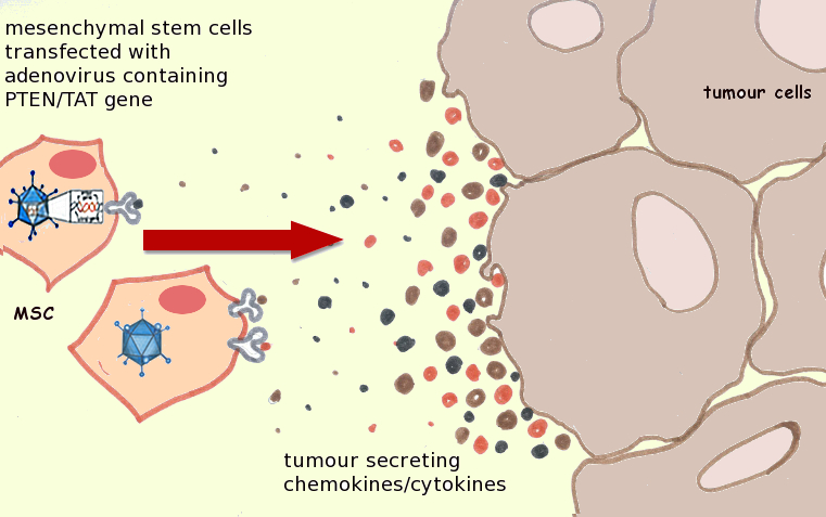
Genetically-modified MSCs home to tumor cells and
accumulate at the tumor site. Image adapted from
http://dx.doi.org/10.1016/j.canlet.2011.02.012.
In the body, MSCs are known to home to sites of acute injury and inflammation and migrate to tumors in response to tumor secretion of growth factors, cytokines, and extracellular matrix (ECM) molecules. However, given their secretion of biomolecules that augment new blood vessel formation, increase inflammation, and degrade the ECM (lending to tumor metastasis), MSCs may promote rather than impede tumor growth and migration, and confounding results from a number of in vitro and in vivo studies have been published to date.  Furthermore, it has been suggested that the ability of MSCs to interact with malignant cells and cancer stem cells might preclude their safe therapeutic application, particularly in patients with dormant or undiagnosed cancers. Despite these concerns, MSCs can serve as an effective ‘Trojan Horse’ for the targeted delivery of anticancer genes, proteins and drugs to tumor cells.  Such targeted delivery can reduce the unsavory systemic side effects that often result from the use of anti-cancer agents, reducing patient morbidity and improving quality of life.  

MSCs can serve as an effective ‘Trojan Horse’ for the targeted delivery of anticancer genes, proteins and drugs to tumor cells.

Recently, a review was published focusing not only on the application of MSCs for the targeted delivery of anti-cancer agents to tumors, but also on the molecular mechanisms of MSC accumulation in tumors, a poorly understood mechanism.  For MSC-based anti-cancer therapies to be effective clinically, these mechanisms must be understood and successfully exploited.  The authors identified several methods to genetically-modify MSCs that resulted in tumor growth inhibition, metastasis suppression, and prolonged survival upon MSC injection in various tumor-laden animal models.  However, in addition to modification with anti-cancer agents, MSCs must be able to accumulate at the site of the tumor for effective cancer eradication.   The authors postulate that increasing the accumulation efficiency of MSCs at tumor sites can effectively target not only primary tumors but also metastatic lesions. 

March 10, 2014

Mesenchymal Stem Cells: The Workhorse of Regenerative Medicine

Prolific Stem Cell and Cell Therapy Blogger Alexey Bersenev (@cells_nnm on Twitter) has recently written a post on his Cell Trials blog titled "Trends in cell therapy clinical trials 2011 – 2013".  If you are interested in Stem Cells and Cell Therapy, it is highly likely that you already know Alexey.  He is one of the most passionate and dedicated technologists in the area.  If you don't know him,  you can follow his activities at the above links, as well as at his Stem Cell Assays blog and among several LinkedIn Discussion groups.  Alexey reported that his own research shows that Cell Therapy Clinical Trials that are listed in international databases have doubled from 2011 to 2013, going from of 161 to 324. Alexey evaluated the data by the split between industry and academia (three academic trials in 2013 for every industry-sponsored trial), by country, by region, as well as by cell type and clinical indication.  His analysis is very important because with his technical background and expertise (Alexey is an MD/PhD researcher who has been doing hands stem cell research or therapeutic cell manufacturing as his day job for several years), if anyone can correctly categorize a trial, it is Alexey.

When tweeting highlights of his Cell Therapy Clinical Trials blog post (above inset) an astute follower of his picked up on the fact that Mesenchymal Stem/Stromal Cell (MSC) trials are out-pacing the other cell types.  This fact is consistent with another often-cited recent manuscript printed in the journal Regenerative Medicine titled "The Global Landscape of Stem Cell Clinical Trials" (free download available here).  This manuscript also creates its own database and analyzes the various types of trials, with the conclusion that "most of the increase (in Cell Therapy Clinical Trials) since 2006 was due to trials using MSCs".

So WHY are MSC trials outpacing other cell sources?  There are several reasons for this.  We believe this is likely due mostly to: