Showing posts with label RoosterBio. Show all posts

September 12, 2018

Building Effective Multi-Year Process Development Programs: Estimating hMSC Lot Size Ranges for Clinical Manufacturing through Commercial Demand – it is all about the assumptions

Authored by Josephine Lembong, Ph.D., Scientist, Product & Process Development & Jon A. Rowley, Ph.D., Founder and Chief Product Officer, RoosterBio Inc.

Jon Rowley, RoosterBio’s Founder and Chief Product Officer, gave a talk at ISCT 2018 in May and had a set of slides about estimating lot size that had a lot of people scrambling to take notes. We thought we would share the content here at the RoosterBio blog with more discussion and make the content broadly available and open for discussion.  

The concept is consistent with all good strategic planning; 
  1. Understand what the future looks like and work backwards,
  2. Create a model using reasonable to conservative assumptions along the way to estimate a range of needs,
  3. Use this model to lay out what the next several years will look like assuming successful achievement of intermediate milestones, and create multi-year programs that are right-sized, with the right technology platform, for the stage of product / clinical development of your company.
This is the type of consultation that we provide in our Process Design and Acceleration business unit at RoosterBio, which we offer to our customers that are incorporating RoosterBio hMSC cell banks and bioprocess media systems into their product and process development efforts. However, we love to share our knowledge with the broader RegenMed Industry and are offering it up here.

Notes to readers: this exercise has the base assumption that this is an Allogenic, Universal Donor manufacturing process [1] and is not applicable to Autologous or patient-specific products. The core logic would still hold when applied to autologous, but at a much smaller scale.

Lot Size Targets Dictate the Production Platform 

The goal of any process development program is to create a right-sized manufacturing process for your immediate business goals, but with future scalability in mind. Understanding the future lot size requirements will help strategically align manufacturing requirements today with commercial scalability while laying a platform foundation to minimize comparability risks.

First Start with the End in Mind – Engage with Marketing

Get with the business team within your company (usually Strategic Marketing and/or Commercial Operations) and request a numbers-driven discussion on which of the multiple indications that a process development program should be built for. There are usually several therapeutic indications of interest, but it is important to realize that a manufacturing process for an ocular indication that has a dose of 1 million cells/dose is very different than a Crohn's disease indication that has a dose size of greater than 1 billion cells/dose [2]. Each therapeutic indication from a company must be treated as a distinct product and target, with a distinct process. Just because the same cell type is used in both therapeutic products does not mean they share a manufacturing process. This is critical to bring clarity to each program.

“A common pitfall that many cell therapy companies fall into is that each therapeutic indication is not addressed as a unique product with a unique manufacturing process”

Once the team is aligned on the target indication (or set of prioritized target indications), then you need to understand what a reasonable peak commercial market demand would be – this is likely to be found in the business model projections that are built for investors and is a good place to start. If there are 500,000 patients a year in your target indication and the business is aiming to capture 20% of them (or treat 100,000 patients/year) at peak commercial demand, then you need a process that will scale with that over time, assuming both clinical and market success. It is a good idea to establish a range. We often like to perform this exercise with a low, middle and high assumptions (such as 30,000 patients as low, 100,000 as the target, and 250,000 patients treated/year if wildly successful). For this article, we will simply focus on the '100,000 patients treated assumption' for our calculations.

Dosing Assumptions: this is one of the hardest parts

Estimating dosing is sometimes the most difficult part. Does the patient need a single dose or multiple doses, and how many cells per dose? In many cases a dose escalation trial has not yet been performed, so some educated guesstimates are required. Taking a low/mid/high range of estimates also works here, and there is sufficient published work related to hMSCs in different indications that you can get close enough for these purposes [2,3]. For this example, we will assume a cardiac indication. There is a good amount of published work on hMSCs for cardiac indication and we can assume a conservative low dose of 25 million cells, a mid-range dose of 50 million, and a high dose of 100 million [4,5]. In this blog post we will perform all calculations with an assumption of 50 million cells per dose.

Calculating Yearly Product Needs at Peak Commercial Demand
  • At peak demand, we aim to produce 100,000 doses per year, with a size of 50 million cells per dose.
  • 100,000 doses per year ≈ 2,000 doses per week
    • Weekly production is not recommended – it is simply unsustainable, so let’s assume one (1) lot production every two (2) weeks, or 25 lots total in a year (which is still a lot for any manufacturing site)
    • Not all lots will be successful – assume a 10% scrap rate, thus 2-3 lots are lost per year, giving us 22-23 lots per year
    • 100,000 doses per year / ~23 lots per year = 4,450 doses/lot 
    • Assume 10% of lot goes to testing (~450 dose), so a final target lot size of 4,900 doses/lot will be needed to be manufactured at peak commercial demand
It is important to remember that it typically takes multiple years for a successful therapeutic to reach peak demand, so it is not necessary to go to market with a manufacturing process capable of meeting peak demand – especially for an early stage field like Regenerative Medicine. We will create estimates for peak demand, but then focus on building a reasonable “go-to-market” lot size and manufacturing process.

Calculating Total Cells to Manufacture that are Required to Fill into the Final Container
  • We will assume 1 dose goes into one container.
  • We assumed a mid-range dose size of 50 million (i.e. 50 million viable cells per vial, post-thaw).
  • Cell Count is a strict QC parameter with a straight forward specification. It is important to point out that often there is a solid safety factor, or overfill, applied to this metric. You never want to fail a lot on cell count after you spent hundreds of thousands of dollars creating the units – so the risk averse will overfill vials until sampling and cell enumeration is an accurate, consistent, and precise method.
    • Assume 15% (worst case) viability drop and 20% recovery loss during cryopreservation (these assumptions should be data driven from your process, and constantly trended and updated during manufacturing).
    • Based on the above assumptions, to consistently achieve 50 million viable cells after thaw, it is required to target filling at 75 million viable cells into the final container:
      • 75 million viable cells – 15 million (20% total cell loss on recovery) = 60 million viable cells 
      • 60 million – 9 million (15% viability drop) = 51 million viable cell target specification
  • NOTE: some programs will only overfill by 10-20%, but we find that with this range it is very difficult to routinely obtain successful cell counts in a manufacturing/QC environment, so we recommend 30-50% overfill for calculations.

Calculating Lot Size Requirements at Peak Commercial Demand

One unfortunate fact of cell manufacturing is that you need to manufacture more cells than you need to fill, since there will be losses associated with the post-harvest processing steps. Many biological manufacturing processes have losses in the 40%-60% range in this “downstream” processing.  For our process, we go through the following math:
  • 75 million cells/dose × 4900 doses per manufacturing lot = 368 billion viable cells needed to target 4900 doses from every manufacturing lot.
  • We assume 30% post-harvest cell loss during processing for this exercise – thus at least 525 billion cells need to be manufactured in order to have 368 billion cells during the fill unit operation.
    • 525 billion viable cells – 157 billion (30% post-harvest cell loss) = 368 billion viable cells 
  • 30% cell loss does seem significant, however every process development group will have a team working to optimize their process to minimize this number.  It is likely that recoveries from earlier processes will be much greater than 30%, and with optimization at large scale it is possible to improve.  For modeling, it is always worth using reasonable to conservative estimates.
In conclusion, we arrived at the following numbers:
  • At peak commercial demand, 23 successful lots of 525 billion cells per lot need to be manufactured in order to meet the demand, assuming a mid-range dose for a cardiac indication aimed to treat 100,000 patients per year. 
This calculation does not take into account further downstream losses associated with stability, multiple distribution sites, safety stocks – which will just make the numbers worse. For this exercise, we will stop here. The 525 billion viable cells per lot is the number we were looking for, and it is this number that will drive the production platform decisions for streamlined hMSC clinical manufacturing.

For an early-stage RegenMed company, a go-to-market lot size of ~20% of the “peak commercial scale” is a reasonable target to plan for, so a ~100 billion cells/lot would be a good target number to develop and run your Phase III trial at.

We’d like to summarize this post with a few number recommendations:


Phase III/Go-to-Market: ~100B cells

Phase II (100-200 patients over 3-4 lots): 15-25B cells – this is an intermediate step up from Phase I and within a log of the Phase III/go-to-market process.

Phase I (small scale): 5-10B cells

The next stage is to lay out the technology platforms for the various manufacturing unit operations that are capable of processing these cell numbers. This will be the topic of our next blog post.

References:
  1. Simaria et al., 2013. Allogeneic cell therapy bioprocess economics and optimization: Single‐use cell expansion technologies. Biotechnol Bioeng 111(1): 69-83. doi: 10.1002/bit.25008 
  2. Olsen et al., 2018. Peak MSC—Are We There Yet? Front Med 5:178. doi: 10.3389/fmed.2018.00178 
  3. Squillaro et al., 2016. Clinical Trials With Mesenchymal Stem Cells: An Update. Cell Transplant 25(5):829-848. doi: 10.3727/096368915X689622 
  4. Majka et al., 2017. Concise Review: Mesenchymal Stem Cells in Cardiovascular Regeneration: Emerging Research Directions and Clinical Applications. Stem Cells Transl Med 6(10):1859-1867. doi: 10.1002/sctm.16-0484 
  5. Golpanian et al., 2016. Concise Review: Review and Perspective of Cell Dosage and Routes of Administration From Preclinical and Clinical Studies of Stem Cell Therapy for Heart Disease. Stem Cells Transl Med 5(2):186-191. doi: 10.5966/sctm.2015-0101


June 16, 2016

At the Cutting Edge of 3D Bioprinting: WBC 2016 Round Up Part II

A guest blog post by RoosterBio Travel Award winner, Ian Kinstlinger.

Presenting on Open-source Selective Laser 
Sintering at WBC2016!
Since 1980, the international community of biomaterials scientists and engineers has convened every four years to discuss the cutting edge of biomaterials research. This year’s 10th World Biomaterials Congress (WBC) brought us to lovely Montreal, Canada for a stimulating week of workshops, talks, posters, and social activities. I was honored to present my work from the Miller Lab at Rice University in both a podium talk and a poster session.

Our lab is broadly interested in developing strategies to construct vascular networks within engineered tissues. In my research, I have developed a platform technology which uses 3D printed carbohydrates as templates around which cells and biomaterials can be assembled. Dissolving the sugar away gives you an engineered tissue with perfusable channels; we believe that these constructs will be useful for understanding the mass transport requirements and emergent properties of engineered living tissue.

An overview of one method our lab has introduced to create 
embedded vascular networks in biomaterials.
I used my poster to spread the word about our lab’s Open-source Selective Laser Sintering technology and my podium talk to describe how we’ve adapted this system to perform laser-based 3D printing of carbohydrate materials. I was thrilled to have a large audience for my talk, followed by several insightful questions. My poster also received a steady stream of visitors, many of whom are involved in the open-source hardware community and were eager to talk about hardware hacking for biomaterials. That work was actually published earlier this year – and RoosterBio hMSCs were absolutely central. Their high quality and robust differentiation response made characterizing biocompatibility of materials quite straightforward.

A couple of key presentations stood out at WBC 2016:

Nano- and Micro-fabricated Hydrogels for Regenerative Engineering
  • Dr. Ali Khademhosseini, Khademhosseini Lab, Harvard University
  •  Dr. Khademhosseini gave an illuminating keynote on the many angles from which his lab is using bioprinting technologies to fabricate functional biological structures. He is also emerging as a leader in the field of integrated organ-on-chip drug screening platforms.

Injection of Dual-Crosslinking Hydrogels to Limit Infarct Induced Left Ventricular Remodeling
  • Dr. Jason Burdick, Polymeric Biomaterials Laboratory, University of Pennsylvania
  •  The Burdick lab has developed an innovative class of supramolecular biomaterials specifically targeted for 3D printing applications. The gels are shear-thinning due to their non-covalent crosslinks, and thus are amenable to extrusion printing. These materials are also useful as injectables for reducing left ventricular remodeling after heart attack.

Photoreversible patterning of hydrogel biomaterials with site-specifically-modified proteins
  • Dr. Cole DeForest,  DeForest Research Group, University of Washington
  •  Much like our lab is interested in patterning biomaterial architecture via 3D printing, the DeForest group is patterning functional proteins into materials through some very clever photochemistries. Their techniques give them spatiotemporal control over the incorporation of various full proteins into synthetic hydrogels.

It was tremendously exciting to see so many investigators working on 3D printing of biomaterials. I counted at least seven sessions devoted to the topic and was also impressed by the low-cost printers and inks now hitting the market, including RoosterBio’s new ready-to-print hMSC products. The diverse hardware and materials that have been introduced in the past few years are already transforming the field! It will be very interesting to see in the coming years whether these new techniques give way to novel insights into cell and tissue function in vitro, as many groups are currently promising.

It is also not yet clear whether the same groups who are mastering the materials and fabrication technology have the resources and expertise to analyze complex biological phenomena in their printed structures. A greater level of collaboration between biologists and materials/fabrication engineers may be necessary in the future to make progress in this area. I am going to end with shameless plug for my recent review article in Lab on a Chip which discusses 3D printing approaches for fabricating vascular networks and addresses the need for increased communication between biologists and materials scientists.

WBC 2016 was an incredible conference in which I got to present my work, learn about key advances in biomaterials, meet leaders in the field, and explore Montreal. Thanks so much to RoosterBio for providing the highest quality hMSCs and for their support of my work through a travel grant! 

June 14, 2016

World Biomaterials Congress 2016 Meeting Round Up Part I: Guest Blog Post

A guest blog post by RoosterBio Travel Award winner, Gisele Calderon.


The World Biomaterials Congress (WBC) takes place every four years with an energy rivaling the Olympics. This Congress is the largest gathering of biomaterials-focused researchers with over 1,200 oral presentations and 2,400 poster presentations representing over 60 countries. I am incredibly grateful to have been given the opportunity to present my work to and learn from the World’s finest leaders of the field.

Our work in the Miller Lab at Rice University focuses on vascularizing engineered tissues to address the metabolic needs of these complex tissues via various techniques. I develop a system to monitor cellular morphogenesis toward a stable capillary plexus allowing biology to dictate the architectural hierarchy. The cell-cell interactions between the endothelial cells derived from an iPS source and human mesenchymal stem cells tend to enhance the stability of the putative capillaries we form. Our novel multicolor genetic reporter system is enabling a new class of longitudinal studies of tubulogenesis and their integration with 3D printed vasculature.

I was overjoyed to present my work to curious peer grad students, esteemed thought-leaders, and industry representatives. I was fortunately located near the coffee so my poster received a high amount of traffic! I particularly enjoyed how accessible all of my science idols were during the Congress. WBC ran an event where discussion and learning were the central mission.  

The Congress highlighted the field’s latest work and how critical using therapeutically relevant cell types (like RoosterBio’s hMSCs) is for successful tissue engineering strategies and forward progress. I especially enjoyed engaging with investigators sharing the following presentations:

3D Tissue Printing
  •    Dr. Jennifer Lewis, Lewis lab, Wyss Institute, Harvard University
  •    How can we directly print human tissue? Their approach utilizes top-down bioprinting elegantly recreating complex vascular geometries.
  •      A recent publication features RoosterBio’s hMSCs!


3D Printing complex scaffolds using Freeform Reversible Embedding of Suspended Hydrogels (FRESH)
  •    Dr. Adam Feinberg, Regenerative Biomaterials & Therapeutics Group, Carnegie Mellon University
  •      They 3DP crazy complex structures in a gel within gel fashion for ubiquitous support in their soft constructs. A fun note - he was inspired by Salvador Dali’s painting where everything droops down without support!
  •       Here’s their FRESH printing paper.


 Hydrogels with continuously variable stiffness defined by dual-color micro-stereolithography
  •             Dr. Neils B Larsen, PolyCell group, Technical University of Denmark
  •    This group constructs 3DP soft constructs using stereolithography techniques in order to incorporate microvessels in their hydrogels. They are able to achieve consistent channels under 200um with tunable elasticity dependent on wavelength and exposure tie of incident light.
  •        More details can be found here.


In reality, there are details of many more presentations that I would love to share here, but I just couldn’t do justice to the high spirit of scientific rigor. Throughout the Congress, I was actively tweeting about all the excellent work. Follow me @g_caldero! And lastly, thank you, RoosterBio, for the awesome cells AND also the travel award support to attend this exciting Congress! 

April 22, 2015

Adipose- and Bone Marrow-Derived hMSCs: What's the Difference?

INTRODUCTION:

Human Mesenchymal Stem/Stromal Cells, or hMSC, are key components of future therapeutics, engineered tissues, and medical devices and are currently in use in over 400 clinical trials (1). Bone marrow-derived MSC (hBM-MSC) have historically been the most widely used hMSC, but hMSC can be isolated from many tissues of the body including fat, umbilical cord blood, dental pulp, Wharton’s jelly, and peripheral blood. In recent years, human Adipose (or fat) tissue-derived MSC (hAD-MSC) are increasingly used in studies due to adipose tissue having a higher frequency of MSC than bone marrow and the relative ease of collection (2). You can find more information on hMSC by following these links:


A common MSC misconception is that MSC isolated from different tissues are equivalent. hAD-MSC and hBM-MSC, and cells from other tissues, can meet the “traditional” ISCT criteria to identify a cell as an MSC (3,4): adherence to plastic, characteristic surface marker expression profiles (positive for CD73, CD90, CD105; negative for CD34, CD45), and trilineage differentiation to fat, bone, and cartilage.  However, there is widespread acceptance that hMSC achieve their biologic and therapeutic effects in vivo by secreting many bioactive molecules (referred to as the hMSC secretome) that moderate a variety of processes including angiogenesis, immunosuppression, and overall “tissue repair” (5). Despite being similar overall, hMSC isolated from adipose and bone marrow display some differences in functional capabilities (2,6). For example, hBM-MSC are more robust in bone and cartilage differentiation than hAD-MSC and hAD-MSC are more efficient at stimulating angiogenesis than hBM-MSC (2,6,7).

We have recently been applying our manufacturing protocols to adipose-derived hMSC (our newest product) and would like to share some of the similarities and differences in function between hBM-MSC and hAD-MSC that we have observed when these cells are cultured in our media systems with our protocols.  Both populations of hMSC have been manufactured using our GMP-compatible and scalable manufacturing processes, with standardized procedures and with rigorous quality control.  By reporting the differential functional characteristics of these hMSC populations, we assist our customers in making more informed choices on the cell type best-suited to their application(s).


METHODS AND EXPERIMENTAL DESIGN:

Materials & Reagents:  Cell culture reagents, excluding RoosterBio materials, were purchased from Life Technologies, chemicals and reagents for kynurenine measurement were from Sigma, and cultureware was from Corning.  Two vials (1 million cells each) of hAD-MSC, representing two donors, were purchased from ZenBio, and used only for comparison. Other cell products used were RoosterBio hMSC products: Bone Marrow-derived MSC (hBM-MSC, part # MSC-001, MSC-003) and Adipose-derived MSC (hAD-MSC, part # MSC-020, MSC-021). Cells were cultured in RoosterBio High Performance Media (part # KT-001) or DMEM + 10% FBS

Methods: All methods for the analyses shown below are documented under RoosterBio’s Quality Control systems.  For more information, please contact us at info@roosterbio.com.  Detailed methods for priming hMSC can be found in a previous blog post here.


RESULTS:

November 6, 2014

Current Bottlenecks in MSC Research: MSC Misconceptions - Part II

 http://andreyev.com.au/wp-content/uploads/Misconceptions.jpg
We blogged recently about Mesenchymal Stem/Stromal Cell (MSC) Misconceptions that are holding the translational cell therapy field back, as identified by Donald Phinney and Luc Sensebé.  Since we have come to market with our own hMSC product lines, we have spoken with hundreds of MSC researchers and engineers, and we have compiled our own set of misconceptions that we think build off of Dr. Phinney’s and Dr. Sensebe’s initial concept.  This blog post is to share some of the market-based feedback that we have received.

To the list that was published in Cytotherapy, we would like to contribute the following list to the conversation:

1. Tracking MSC passage number is an accurate and reliable means of tracking cell age and standardizing experimental workflow
In many research laboratory environments, cellular age is most often tracked by the number of times a cell has been passaged; however, Passage Number is quite imprecise and not very acceptable as one gets into regulated environments such as translational clinical activities.  It is generally accepted that tracking the Population Doubling Level (PDL) or Cumulative Population Doublings (CPD) of primary cells is a best practice on understanding cellular age in vitro Since it is well documented that PDL impacts hMSC function (see here, here and here), in order to drive consistency into experiments, it has become a best practice to perform experiments or develop products with cells in a consistent range of population doublings where the cell function of interest is still robust.  Furthermore, regulatory agencies are beginning to require reporting of PDLs, or at least cell seeding and harvest densities, for primary cells intended for therapeutic use.  In an effort to drive adoption of PDL tracking and reporting, we’ve created a Best Practices Educational Powerpoint, and free, easy-to-use PDL calculator worksheet we’re happy to share with colleagues.  For your copy, just email us at info@roosterbio.com or subscribe to our blog!

2. Performing experiments with one MSC donor and/or lot is adequate for publication and moving forward with pre-clinical studies
Despite indications of clinical effectiveness of MSCs, there is repeated news of the failure of high-profile MSC trials to demonstrate efficacy in a number of therapeutic applications.  It has been suggested that the large amount of intra- and inter-donor variability in the MSC populations used in these trials may be responsible for their falling short of expectations despite highly encouraging in vitro and in vivo pre-clinical data.  Thus, to ensure the robust production of functional MSC products over a range of applications, experiments should be conducted and systems validated with MSCs from several donors It has been reported that best practices to qualify a manufacturing process should include “at least 3-5 donors”, and it is likely that proper Validation will require many more, and that donor selection may be required (i.e. not every donor will work in the manufacturing process). This is why we, at RoosterBio, believe in providing a number of donor MSC lots, ranging in age and sex, for use in our customer’s research and development experiments.

3. MSCs accelerate cancer…..MSCs can combat cancer

August 25, 2014

Best Practices in MSC R&D: Addressing Donor Variability within your Experimental System

Human MSCs are the single most used cell source for tissue engineering and regenerative medicine applications, and clinical trials involving hMSCs have outpaced all other cell types in recent years (see here and here).  However, despite indications of clinical effectiveness (see here and here), there is repeated news of the failure of high-profile MSC trials to demonstrate efficacy in a number of therapeutic applications (see here, here, here, here and here).  It has been suggested that the large amount of intra- and inter-donor variability in the MSC populations used in these trials may be responsible for their falling short of expectations despite highly encouraging in vitro and in vivo pre-clinical data.

A team led by Steve Bauer at the US FDA has reported that large variations in proliferation, morphology, differentiation capacity, and cell surface marker expression profiles exist within any population of MSCs and that these intra-population heterogeneities may arise as a result of long-term in vitro culture and the in vivo microenvironment (Free article available here.)  In addition, their work has demonstrated that there are inherent differences in MSCs from donors of similar age, and they have noted the “potential for other donor-related factors in MSC biological variability, which may play a role in their clinical usefulness or performance in various model systems.” Other research groups have also corroborated donor-related differences in MSC function, including in response to stimuli, such as challenge with inflammatory cytokines (see here and here).  A review article on developing cell therapy manufacturing processes reinforces that several donors should be tested prior to implementing; 1) changes in media composition (such as serum reduction/elimination or addition of growth supplements), 2) extensions of the product dose population doubling level (PDL), or 3) changes in lot size during scale-up.

March 14, 2014

Using MSCs to Beat Cancer: The Next Big MSC Application

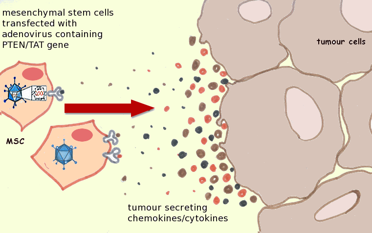
Genetically-modified MSCs home to tumor cells and
accumulate at the tumor site. Image adapted from
http://dx.doi.org/10.1016/j.canlet.2011.02.012.
In the body, MSCs are known to home to sites of acute injury and inflammation and migrate to tumors in response to tumor secretion of growth factors, cytokines, and extracellular matrix (ECM) molecules. However, given their secretion of biomolecules that augment new blood vessel formation, increase inflammation, and degrade the ECM (lending to tumor metastasis), MSCs may promote rather than impede tumor growth and migration, and confounding results from a number of in vitro and in vivo studies have been published to date.  Furthermore, it has been suggested that the ability of MSCs to interact with malignant cells and cancer stem cells might preclude their safe therapeutic application, particularly in patients with dormant or undiagnosed cancers. Despite these concerns, MSCs can serve as an effective ‘Trojan Horse’ for the targeted delivery of anticancer genes, proteins and drugs to tumor cells.  Such targeted delivery can reduce the unsavory systemic side effects that often result from the use of anti-cancer agents, reducing patient morbidity and improving quality of life.  

MSCs can serve as an effective ‘Trojan Horse’ for the targeted delivery of anticancer genes, proteins and drugs to tumor cells.

Recently, a review was published focusing not only on the application of MSCs for the targeted delivery of anti-cancer agents to tumors, but also on the molecular mechanisms of MSC accumulation in tumors, a poorly understood mechanism.  For MSC-based anti-cancer therapies to be effective clinically, these mechanisms must be understood and successfully exploited.  The authors identified several methods to genetically-modify MSCs that resulted in tumor growth inhibition, metastasis suppression, and prolonged survival upon MSC injection in various tumor-laden animal models.  However, in addition to modification with anti-cancer agents, MSCs must be able to accumulate at the site of the tumor for effective cancer eradication.   The authors postulate that increasing the accumulation efficiency of MSCs at tumor sites can effectively target not only primary tumors but also metastatic lesions. 

March 10, 2014

Mesenchymal Stem Cells: The Workhorse of Regenerative Medicine

Prolific Stem Cell and Cell Therapy Blogger Alexey Bersenev (@cells_nnm on Twitter) has recently written a post on his Cell Trials blog titled "Trends in cell therapy clinical trials 2011 – 2013".  If you are interested in Stem Cells and Cell Therapy, it is highly likely that you already know Alexey.  He is one of the most passionate and dedicated technologists in the area.  If you don't know him,  you can follow his activities at the above links, as well as at his Stem Cell Assays blog and among several LinkedIn Discussion groups.  Alexey reported that his own research shows that Cell Therapy Clinical Trials that are listed in international databases have doubled from 2011 to 2013, going from of 161 to 324. Alexey evaluated the data by the split between industry and academia (three academic trials in 2013 for every industry-sponsored trial), by country, by region, as well as by cell type and clinical indication.  His analysis is very important because with his technical background and expertise (Alexey is an MD/PhD researcher who has been doing hands stem cell research or therapeutic cell manufacturing as his day job for several years), if anyone can correctly categorize a trial, it is Alexey.

When tweeting highlights of his Cell Therapy Clinical Trials blog post (above inset) an astute follower of his picked up on the fact that Mesenchymal Stem/Stromal Cell (MSC) trials are out-pacing the other cell types.  This fact is consistent with another often-cited recent manuscript printed in the journal Regenerative Medicine titled "The Global Landscape of Stem Cell Clinical Trials" (free download available here).  This manuscript also creates its own database and analyzes the various types of trials, with the conclusion that "most of the increase (in Cell Therapy Clinical Trials) since 2006 was due to trials using MSCs".

So WHY are MSC trials outpacing other cell sources?  There are several reasons for this.  We believe this is likely due mostly to:

February 26, 2014

Democratizing Living Cellular Technology

@JennWebb recently wrote an article for the O’Reilly Radar titled Democratizing Technology and the Road to Empowerment.  She starts out the article with a nice summary of what it means to Democratize Technology.  Jenn writes “Advancements in technology are making what once was relegated only to highly educated scientists, engineers and developers accessible to — and affordable for — the mainstream.“  Now, the blog she writes for is focused on the intersection of Hardware and Software (or the “physical and digital worlds” is how they phrase it), while we at RoosterBio are imagining a World where biotechnology, specifically living cellular technologies, are simplified and cost-reduced to the point that you don’t have to be a PhD researcher in a well-funded laboratory to perform your own experiments or build novel things out of living cells. The concept of biology paralleling the advances of IT are well laid out elsewhere.

Today, it is much easier to incorporate living cells into your research than it was 20 years ago.  This is evidenced by the proliferation of Cell Biology capabilities in Engineering departments all over the world as Biomedical Engineering has turned into a formalized academic discipline.  When I was doing undergraduate research at the University of Michigan in the early 1990’s, it took months and several collaboration attempts before we could get living cells onto the biomaterial constructs we were making at the time.  Today, it is more commonplace to find the tools to marry the Worlds of Cell Biology and Engineering in the same laboratory.  Despite this, the total number of labs with such capabilities and expertise is still very small.

 We believe that the steps required to fully Democratize Cellular Technologies will be to:

February 20, 2014

Current Bottlenecks in MSC Research

Mesenchymal Stem Cells (MSCs) are widely studied in academic circles and an attractive cell source for clinical applications. MSCs not only possess the ability to self-renew and differentiate to a number of mesenchymal lineages in vitro and in vivo [1,2], but these cells also secrete a cadre of potent trophic factors that contribute to tissue remodeling and modulate the host immune response, making them an attractive cellular biopharmaceutical for the treatment of a number of degenerative diseases and traumatic injuries [3]). However, there is a significant need to improve current methods to efficiently expand standardized, well-characterized MSCs in vitro to the cell numbers needed for widespread, off-the-shelf clinical use

Despite their immense therapeutic potential, MSCs are very rare, comprising only 0.001%-0.01% of the mononuclear cells in the bone marrow [1]. Since a typical adult bone marrow aspirate yields very few MSCs (roughly 1 out of every 10,000 cells) [4] prolonged in vitro expansion is typically necessary before clinical use. However, MSCs will often senesce (i.e. stop growing) in culture before adequate cell numbers for transplantation (on the order of a billion cells) can be obtained. In addition, prolonged in vitro culture of MSCs has been shown to diminish their multilineage potential and impair their immunosuppressive activity [5-8]. The aforementioned challenges associated with MSC culture currently limit their therapeutic potential, and a significant need remains for methods to efficiently expand multipotent MSCs ex vivo.  Therefore, extensive effort has been put into developing methods to expand MSCs while maintaining their differentiation potential and paracrine activity. Cell plating density, culture surfaces, and the addition of growth factor supplements have all been investigated. Of these variables, the use of growth factor and cytokine supplements has proven to effectively modulate MSC growth and self-renewal [9] while maintaining desirable cell characteristics.   

There are three major challenges that cell and tissue engineering technology efforts face today. These challenges are:

February 12, 2014

Welcome to the RoosterBio Blog

Everyone at RoosterBio is extremely excited to be launching our company, shipping our first products, and interacting with tissue engineers, cell therapists, synthetic biologists, and our customers to learn about innovative research in these fields, share our knowledge, and contribute to the Cell and Tissue Engineering Revolution. The central theme behind RoosterBio, or what we call our “business hypothesis”, is that as living cellular technologies become more affordable, easier to access, and much simpler to incorporate into product development efforts– there will be a rapid acceleration in products coming to market that incorporate these technologies. We believe that we can help shape this new market by Democratizing Cell Technologies and making them abundant, affordable, and much simpler to translate into the clinic.

We have assembled a team at RoosterBio that have years of experience in stem cell R&D, cell therapeutic product and process development, and manufacturing operations, and with this blog we hope to share many of the topics that we are so enthusiastic about.  We gravitate toward technologies at the technical interfaces of fields such as 3D printing and tissue engineering; thus 3D bioprinting of tissues will be a big topic on this blog.  We will be sharing our thoughts on step-changes in technology on topics like stem cell therapies, engineered tissues and organs, biological robots, manufacturing technologies, and synthetic biology.  We will also engage in educational posts related to our expertise, and comment on broad themes that are facing the industries that we care about, such as establishing standards in the stem cell arena.  The developments in progressive fields always have profound effects on science and society, and we believe we are on the verge of the cell and tissue engineering revolution. 

This blog is targeted at the scientists, technologists, engineers, and doctors that are working passionately and feverishly to bring cell-based products to patients, as well as for general audiences that are interested in the driving forces behind cellular therapies, regenerative medicines, and tissue engineering technologies.  We hope you will come back to learn and engage in the conversation, and also be a part of the next technology revolution as Biology becomes Technology

We encourage everyone to leave comments, and to feel free to say what is on your mind. We look forward to the dialogue and helping to accelerate the Cell-based BioEconomy!


- The RoosterBio Team